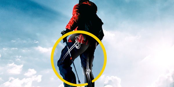
The hip is a ball and socket joint with the ball being formed by the head of the femur (thigh bone) and the pelvic bone forming the socket (acetabulum). The correct alignment of the femur in relation to the pelvic bone plays an important role in the proper functioning and health of the joint. Fractures, non-union of treated fractures or deformities may result in an improper relationship between these structures, leading to wear and tear (arthritis) and pain at the joint, and gait deformities. A femoral osteotomy is a surgical procedure that involves reshaping the upper part of the femur to realign it within the socket. The correction of the hip anatomy reduces the symptoms and progression of arthritis.
Before performing a femoral osteotomy, your doctor will conduct a physical examination and obtain appropriate imaging studies to identify the deformity and decide on the type of osteotomy. During the procedure, your doctor cuts the femur and realigns it correcting rotational or angular deformities. When displacement is severe, the femur may need to be shortened to prevent stress on the surrounding tissues. Metal pins, plates or rods are placed to stabilize the osteotomy site until it heals.
As with all surgical procedures, femoral osteotomy may be associated with certain complications such as infection, nerve and blood vessel injury, inability to achieve complete correction and persistence of pain and non-union.
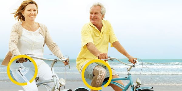
High tibial osteotomy is a surgical procedure performed to relieve pressure on the damaged site of an arthritic knee joint.
It is usually performed in arthritic conditions affecting only one side of your knee and the aim is to take pressure off the damaged area and shift it to the other side of your knee with healthy cartilage. During the surgery, your surgeon will remove or add a wedge of bone either below or above the knee joint depending on the site of arthritic damage.
High tibial osteotomy is commonly used for patients with osteoarthritis that is isolated to a single compartment (unicompartmental osteoarthritis). It is also performed for treating a variety of knee conditions such as gonarthrosis with varus or valgus malalignment, osteochondritis dissecans, osteonecrosis, posterolateral instability, and chondral resurfacing.
The goal of the surgery is to release the involved joint compartment by correcting the malalignment of the tibia and to maintain the joint line perpendicular to the mechanical axis of the leg. There are two techniques that may be used: closing wedge osteotomy and opening wedge osteotomy. The surgeon determines the choice of the technique based on the requirement of the patient.
Closing wedge osteotomy is the most commonly used technique to perform high tibial osteotomy. In this procedure, the surgeon makes an incision in front of the knee and removes a small wedge of bone from the upper part of the tibia or shin bone. This manipulation brings the bones together and fills the space left by the removed bone. The surgeon then uses plates and screws to bind the bones together while the osteotomy heals. This procedure unloads the pressure off the damaged joint area and helps to transfer some of the weight to the outer part of the knee, where the cartilage is still intact.
In this procedure, the surgeon makes an incision in front of the knee, just below the knee cap and makes a wedge-shaped cut in the bone. Bone graft is used to fill the space of the wedge-shaped opening and if required plates and screws can be attached to further support the surgical site during the healing process. This realignment increases the angle of the knee to relieve the painful symptoms.
Complications following high tibial osteotomy may include infection, skin necrosis, non-union (failure of the bones to heal), nerve injury, blood vessel injury, failure to correct the varus deformity, compartment syndrome and deep vein thrombosis or blood clots.

Bethesda Hospital
25 Queenslea Drive, Claremont WA 6010
Tel : 08 9230 633308 9230 6333Fax : 08 9230 6332
Murdoch Square
Suite 206 Level 2 Tower C 44 Barry Marshall Parade, Murdoch WA 6150
Tel : 08 9230 633308 9230 6333Fax : 08 9230 6332
Mandurah/Peel Health Campus
110 Lakes Road Mandurah WA 6210
Tel : 08 9230 633308 9230 6333Fax : 08 9230 6332
Waikiki Specialist Centre
221 Willmott Drive Waikiki WA 6169
Tel : 08 9230 633308 9230 6333Fax : 08 9230 6332
Coastal Orthopaedics-West Coast Health & High Performance
Mineral Resources Park, 42 Bishopsgate St
Lathlain 6100
Tel : 08 9230 633308 9230 6333Fax : 08 9230 6332
Esperance Hospital
Hicks St,
Esperance WA 6450
Tel : 08 9230 633308 9230 6333Fax : 08 9230 6332
Dunsborough Sports Medicine Centre
4A Turnberry Lane Dunsborough WA 6281
Tel : 08 9230 633308 9230 6333Fax : 08 9230 6332
Kalgoorlie Coastal Orthopaedics
Unit 2, 102 Brookman Street
Kalgoorlie WA
Tel : 08 9230 633308 9230 6333Fax : 08 9230 6332
St John of God Mount Lawley Medical Centre
Suite 4 Level 2,
Ellesmere Rd,
Mount Lawley WA 6050
Tel : 08 9230 633308 9230 6333Fax : 08 9230 6332